
Fig. 8.
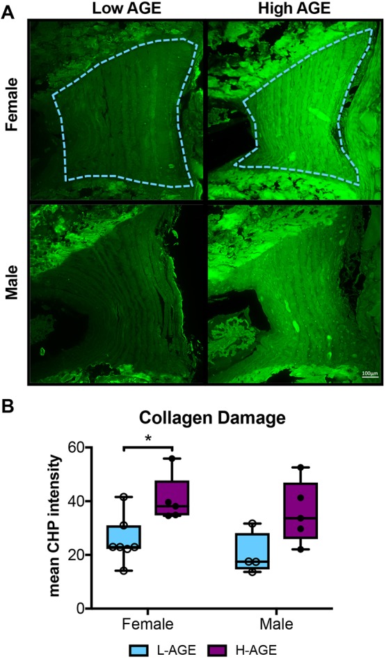
Molecular assessment of collagen. (A) CHP-stained fluorescent images of anterior AF exhibit increased CHP staining in H-AGE compared to L-AGE in females (‘F’) and males (‘M’). The region of interest is outlined in blue. (B) Quantification of CHP mean intensity shows significant collagen damage in H-AGE F (n=5) compared to L-AGE F (n=7). No differences were detected in between males (L-AGE M n=4, H-AGE M n=5). Data are presented as box plots from minimum to maximum±s.d. P-values are based on two-tailed unpaired Student's t-test with Bonferroni correction and significant if P≤0.05 (*).