Fig. 6.
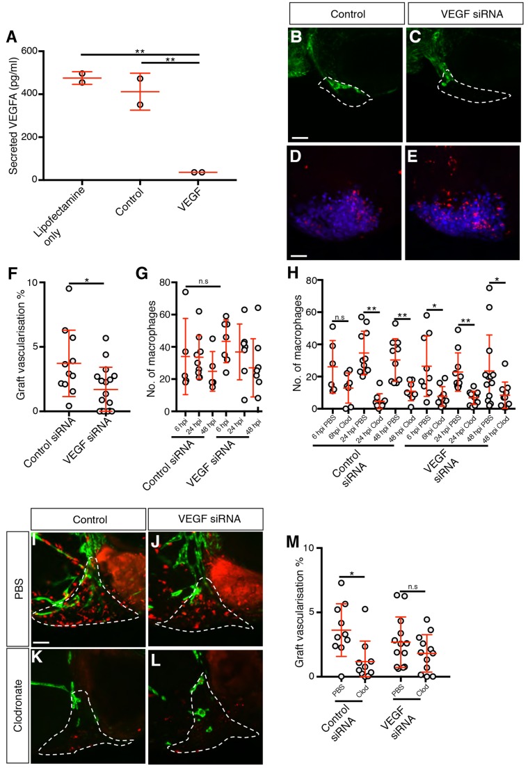
Macrophages are not required for vascularisation in MDA-MB-231 xenografts depleted of VEGFA. (A) Quantitation of secreted VEGFA levels in 2×10⁵ siRNA-treated cells, n=2. (B,C) Confocal images taken at 2 dpi of kdrl:EGFP-expressing vessels (green) in zebrafish embryos implanted with MDA-MB-231 xenografts (white dashed line) transfected with either control (B) or VEGFA siRNA (C). (D,E) Confocal images taken at 6 hpi of mpeg1:mCherry-expressing macrophages (red) and MDA-MB-231 xenografts (blue). (F,G) Quantitation of graft vascularisation at 2 dpi, n>10 (F), and of graft-associated macrophages at 6, 24 and 48 hpi, n>4 (G). (H) Quantitation of graft-associated macrophages at 6, 24 and 48 hpi, in embryos injected with either PBS-containing or clodronate-containing liposomes, n>5. (I-L) Confocal images taken at 2 dpi of mpeg1:mCherry-expressing macrophages (red) and kdrl:EGFP-expressing blood vessels (green) in zebrafish embryos implanted with MDA-MB-231 xenografts (white dashed lines) transfected with either control (I,K) or VEGFA (J,L) siRNA that have been injected with either PBS-containing liposomes (I,J) or clodronate-containing liposomes (K,L). (M) Quantitation of graft vascularisation at 2 dpi in embryos injected with either PBS-containing or clodronate-containing liposomes, n>9. Error bars represent s.d. n.s, P>0.05; *P<0.05; **P<0.01 by either one-way ANOVA (A) or t-test (F-H,M). Scale bars: 50 µm.

