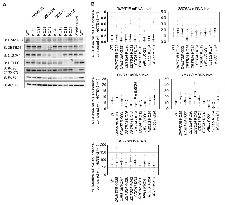
Figure 2. Establishment of cells carrying mutations in ICF genes and Ku80.
(A) Representative blots from biological triplicate showing expression of DNMT3B, ZBTB24, CDCA7, HELLS, and Ku80 proteins in WT and mutant HEK293 cells. ACTB was an internal control. Null mutant cells were designated knockout (KO, plus clone number), and a Ku80 hypomorphic mutant was designated mut24. The overall structure of each protein and regions recognized by the antibody (immunogen) are shown in Supplemental Figure 1. Full Western blot images show that no truncated proteins are present (Supplemental Figure 3). To detect Ku80, rabbit monoclonal EPR3467 antibody was used. (B) Levels of mRNAs in the mutant cells were measured by real-time qPCR in biological triplicate and technical triplicate (n = 9). The mRNA abundance (ΔCt) of each gene was calculated in comparison with that of ACTB and is shown as percent relative expression (2–ΔCt × 100). Data are mean ± SEM. *P < 0.0011 and **P < 0.0002 (Student’s 2-tailed t test) were considered statistically significant at the 5% and 1% levels, respectively, after Bonferroni correction. The exact P value, which was significant (P < 0.01) before the correction, is shown for reference.

