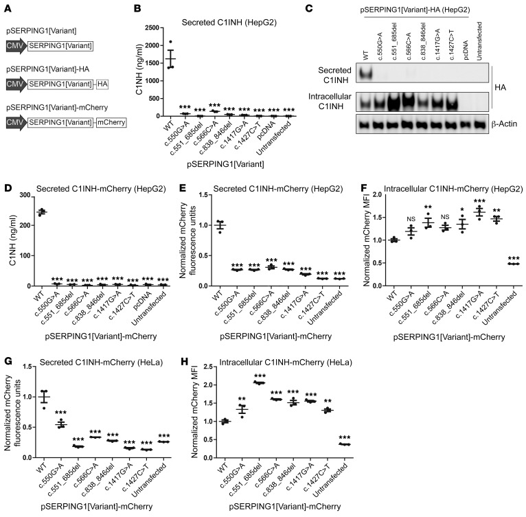
Figure 2. Expression and severely reduced secretion of C1INH encoded by the 6 studied SERPING1 gene variants.
(A) Schematic representation of the vector types used in this study to express SERPING1 gene variants. Black arrows indicate the promoter derived from cytomegalovirus (CMV). (B) C1INH levels in medium from HepG2 cells transfected with 900 ng pSERPING1[Variant] measured by ELISA. (C) Western blot analysis of medium protein and total protein derived from HepG2 cells transfected with 900 ng pSERPING1[Variant]-HA. The secreted and intracellular C1INH levels were detected using a HA-specific antibody. (D) C1INH levels in medium from HepG2 cells transfected with 900 ng pSERPING1[Variant]-mCherry measured by ELISA. (E and F) Secreted and intracellular levels of C1INH in HepG2 cells measured by mCherry fluorescence intensity. (G and H) Secreted and intracellular levels of C1INH in HeLa cells measured by mCherry fluorescence intensity. Cells were transfected with different pSERPING1[Variant]-mCherry vectors. The amount of C1INH-mCherry secreted into the medium was determined using a fluorescence scanner (E and G) and the intracellular level by flow cytometry (F and H). (B–H) Transfections were carried out in triplicate (n = 3) and similar results were seen in at least 2 independent experiments. Data are mean ± SEM. *P < 0.05, **P < 0.01, ***P < 0.001, compared with WT. Statistical analyses were performed by 1-way ANOVA with Dunnett’s multiple comparison test. MFI, median fluorescence intensity.

