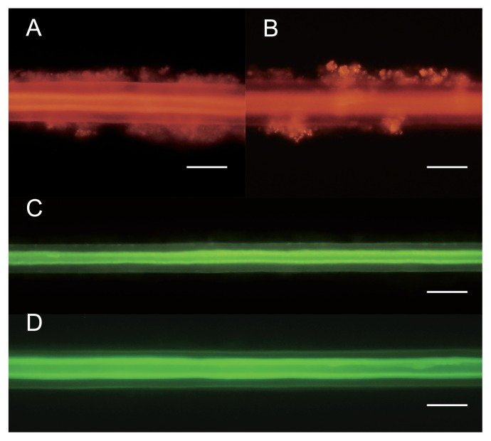
Fig. 2.
Fluorescence microscopy of setae of S. crosnieri individuals during methane-fed rearing. Fluorescence microscopy was performed for the setae of S. crosnieri after rearing for 3 months (A and C) and 12 months (B and D). FISH with the MEG2 probe specifically detects Methylococcaceae-affiliated epibionts on the setae (A and B). FISH with the EPI653 probe specifically detects Sulfurovum-affiliated epibionts on setae (C and D). Scale bars indicate 20 μm (A and B) and 30 μm (C and D).

