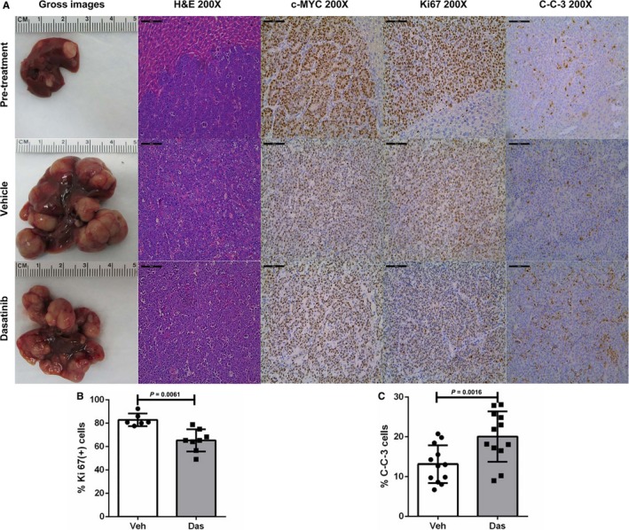
Figure 4.

Dasatinib treatment inhibits proliferation and promotes apoptosis in c‐Myc mouse HCC. A, Gross images, H&E staining and immunohistochemical staining of pretreated, vehicle treated, and Dasatinib treated FVB/N mice. Scale bars: 100 μm for H&E, c‐Myc, Ki67 and C‐C‐3 staining. B, Quantification of Ki67 immunostaining. Each dot represents one measurement replicate (Veh, n = 6; Das, n = 8). C, C‐C‐3 apoptosis upon Dasatinib treatment. Each dot represents one measurement replicate (Veh, n = 12; Das, n = 12). Data are presented as mean ± SD; and P‐value was calculated using Mann‐Whitney U test. C‐C‐3, Cleaved Caspase 3; Das, Dasatinib; SL, surrounding liver; T, tumor; Veh, Vehicle
