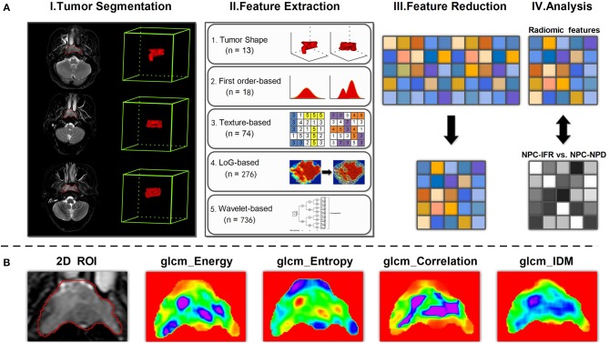
Figure 3.
(A) Workflow of radiomic analysis for discrimination between NPC-IFR (NPC with in-field recurrence) and NPC-NPD (NPC with non-progression disease). I, Image segmentation was performed on SPAIR T2W MR images. II, Features were extracted from the tumor contours on the MR images using shape, first order, texture, LoG and wavelet-based method. III, Principal component analysis (PCA) was performed on significant features for dimension reduction. IV, For the analysis, principal components derived from significant features were combined with supervised machine learning method for prediction of NPC-IFR vs. NPC-NPD. (B) Examples of feature maps computed from two-dimensional tumor region by using GLCM method (e.g., Energy, Entropy, Correlation, InverseDifferenceMoment [IDM]).

