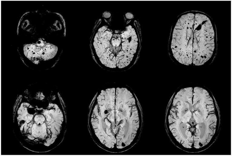
Figure 2.
CCM lesions in Family One detected by SWI-MRI. The proband's SWIs (II:1) revealed dot and patchy low intensity lesions on temporal lobe, cerebellum and thalamus (upper row). SWI (II:2) showed 2 popcorn-like lesions in left occipital region and right temporal pole, which were very dark, suggesting chronic bleeding.

