FIGURE 2.

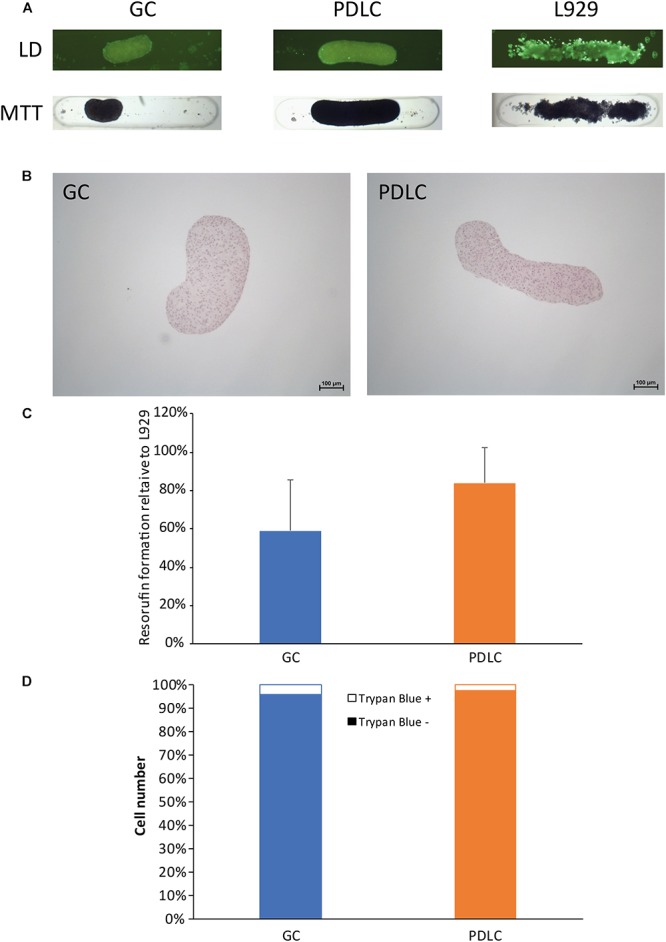
Rods maintain viability based on live/dead (LD) staining, MTT staining, histological analysis, and resazurin-based toxicity assay, and cell count. To assess the viability of the rod microtissues live/dead staining, the MTT staining (A), and histological evaluation by hematoxylin- eosin staining (B) were performed with rods after 24 h. Furthermore resazurin-based toxicity assays (C), and trypan exclusion assay (D) were done. Experiments were performed three times with four replicates each. (C) Bars represent mean ± standard deviation relative to L-929 cells. Gingiva-derived cells (GC); periodontal ligament-derived cells (PDLC).
