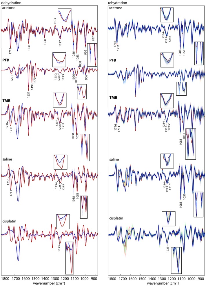
Figure 6.
IR Average spectra (second derivative) of ssDNA treated with acetone (control 1) PFB, TMB, saline (control 2) and cisplatin in the course of dehydration (left) colour-coded from blue (hydrated) to red (dehydrated) and in the course of rehydration (right) colour-coded from red (dehydrated) to blue (hydrated). The inserts show spectral features around 1225 and 1088/1051 cm−1.

