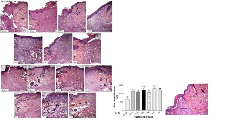
Figure 2a: Haematoxylin and eosin (H&E) stained sections on the 8th day of treatment at 4X magnification showing the area of healing at the site of excision. All the treated groups showed better healing compared to control.
Figure 2b: Haematoxylin and eosin (H&E) stained sections on the 16th day of treatment at 4X magnification showing the area of healing at the site of excision. Better tissue remodeling was observed in all the treatment groups compared to control. Honey, GG, and Tot also showed the growth of hair follicles (yellow arrow). *indicates the healing at the site of the wound.
Figure 2c: Graphical representation of the rate of epithelialisation as quantified histologically in all the treated groups compared to control. *p<0.05 vs control, **p<0.01 vs control.
Figure 2d: Representative image showing the demarcation between the normal and the healing skin (yellow dotted line). Neo-epithelization was measured from the point of demarcation.

