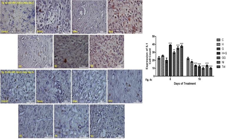
Figure 4a: IHC-HRP staining IL1 beta (stained red) in the granulation tissue of the healing rat skin on the 8th day of treatment at 40X magnification.
Figure 4b: IHC-HRP staining IL1 beta (stained red) in the granulation tissue of the healing rat skin on the 16th day of treatment at 40X magnification.
Figure 4c: Graphical representation of the cell count showing inflammatory responses (IL1-beta expression) on day 8 & 16 in treated groups compared to control. *p<0.05 vs control, **p<0.01 vs control, ***p<0.001 vs control.

