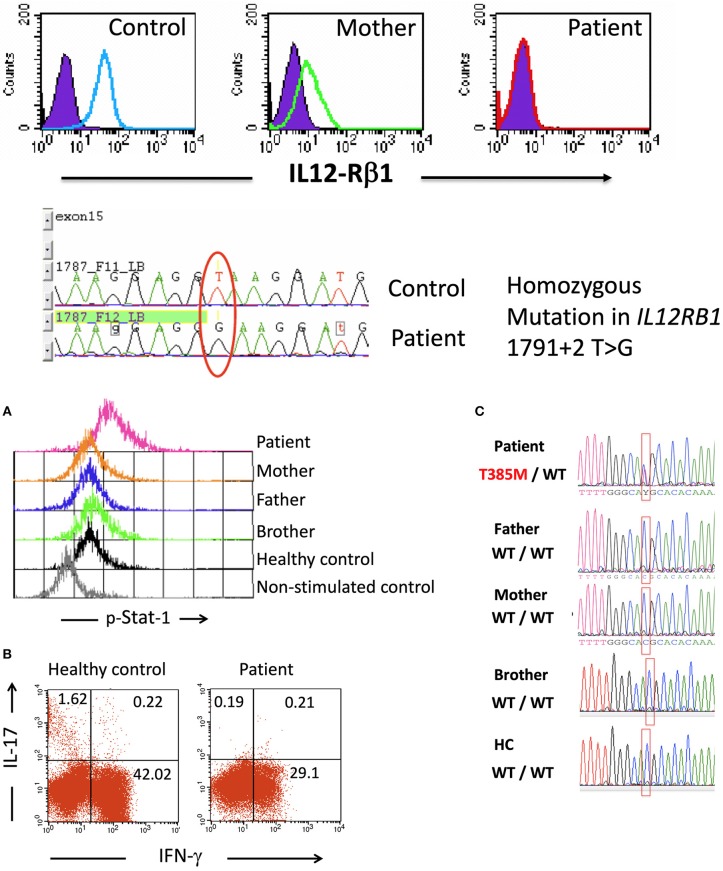
Figure 1.
Upper: IL-12Rβ1 expression on day 3 PHA-T blasts in cells from a healthy control, the mother of patient 1, and patient 1 as assessed by flow cytometry (filled histograms, isotype internal control, and open histograms, β1 chain of IL-12 receptor). Compared to a healthy control, the DNA sequencing showed a mutation at exon 15—intron 15–16 in the samples from the patient. Lower (A) Intracellular p-Stat1 in monocytes from patient 2 and controls assessed by flow cytometry showing Stat1 hyperphosphorylation in monocytes from the patient (peripheral blood mononuclear cells, PBMCs, were stimulated with recombinant human IFN-γ for 30 min; then, the membranes were labeled with anti-CD14+, and the cells were intracellularly stained for p-Stat1). (B) Intracellular production of IFN-γ and IL-17 in CD3+ blood cells from a control and patient 2. PBMCs were stimulated with PMA + ionomycin for 6 h in the presence of Brefeldin A, labeled on membranes with anti-CD3 PerCP and intracellularly stained with anti-IFN-γ Alexa Fluor 488 and anti-IL-17 PE. The numbers shown in the quadrants represent the percentages of positive cells. (C) Sequencing of the STAT1 gene showed the heterozygous mutation T385M in patient 2, and this mutation was absent from her parents, brother and healthy controls. Lower panel (C) is taken from (21), with permission.

