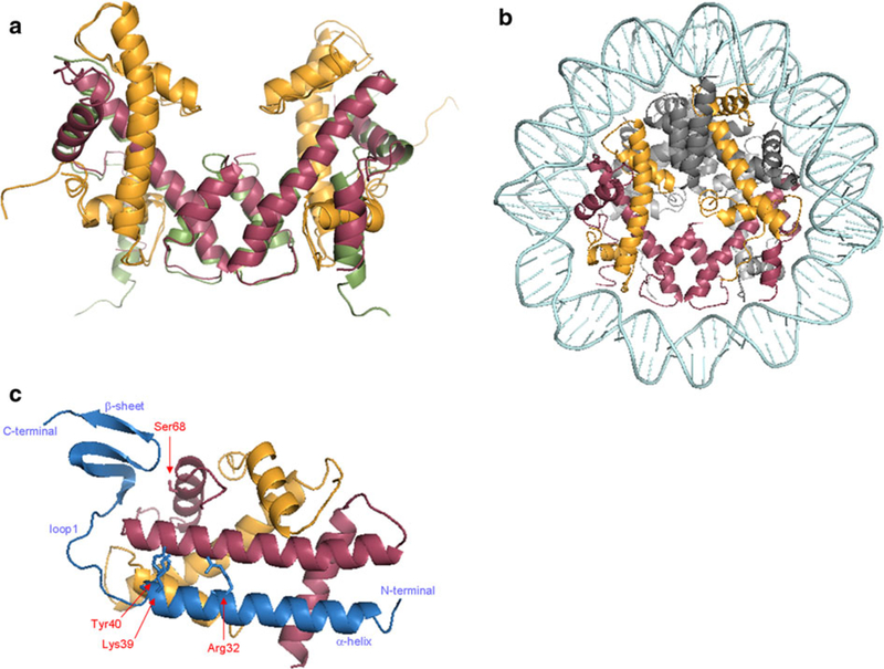
Fig. 2.

Crystal structure of human CENP-A complexes. a Superimposition of the (CENP-A-H4)2 tetramer (PDB 3NQJ; Sekulic et al. 2010) and H3 (PDB 1AOI; Luger et al. 1997). b View in the axis of the DNA helix of the CENP-A octamer (PDB 3AN2; Tachiwana et al. 2011). c Structure of the HJURP-CENP-A-H4 trimer (PDB 3R45; Hu et al. 2011). Important residues for the interaction between these three proteins are localized: Serine68 (Ser68) on CENP-A, Arginine32 (Arg32), Lysine39 (Lys39), and Tyrosine40 (Tyr40) on HJURP. All ribbon diagrams were generated in Pymol (CENP-A in red, H4 in orange, H3 in green, H2A and H2B in gray, and HJURP in blue)
