
Fig. 4.
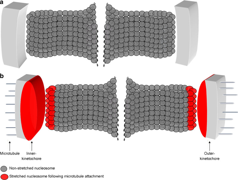
Deformation of the inner kinetochore after microtubule attachment during mitosis. a For the correct chromosome segregation, kinetochore is formed at the centromeric locus. b The correct attachment of microtubules to the outer kinetochore leads to produce a tension, which will trigger structural changes of the inner kinetochore (oval red structure in b), but not the outer kinetochore (gray rectangle in b). The stretch of the inner kinetochore involved constitutive centromere-associated network (CCAN) proteins, composed for example of CENP-A, CENP-C, CENP-N, CENP-T, CENP-W, CENP-S, or CENP-X