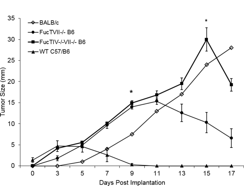
Figure 4: J558L tumors showed early growth in skin of BALB/c and FucTVII−/− B6 mice but not WT B6 mice; early tumor growth was even greater in skin of double-knockout FucTIV−/−/VII−/− B6 mice than in single knockout FucTVII−/− B6 mice.

J558L cells were injected into abdominal skin and external dimensions of the resulting tumors were measured. Tumors grew progressively in the skin of syngeneic BALB/c mice (n=1) and allogeneic FucTVII−/− B6 mice (n=7) for the first 9–11 days. Tumors grew faster in double knockout FucTIV−/−/VII−/− B6 mice (n=11) than either BALB/c or single knockout FucTVII−/− B6 mice (n=5) (day 15, t = 5.31, p < 0.001). Tumors began to regress later in the double knockout FucTIV−/−/VII−/− B6 mice versus the single knockout FucTIV−/−/VII−/− B6 mice. In a separate experiment, J558L tumor cells injected into abdominal skin of allogeneic WT B6 mice (n=5) did not show significant growth. Of note, no tumor growth was seen in >15 WT B6 mice. Comparisons of mean tumor size at day 9 show tumor size was significantly larger in single knockout FucTVII−/− B6 mice (n=5) (t = 22.4, p < 0.001) and double knockout FucTIV−/−/VII−/− B6 mice (n=11) (t = 21.8, p < 0.001) than in WT B6 (n = 5). Comparisons of mean tumor size at day 15 show tumors were significantly larger in double knockout FucTIV−/−/VII−/− B6 mice (n=11) than single knockout FucTVII−/− B6 mice (n=5) (t = 22.4, p < 0.001). All tests are 2 tailed t-tests. The bars represent the mean ± SE. These tumor measurements were taken in vivo and include surrounding skin and associated inflammatory infiltrates, so tumor sizes may be overstated.
