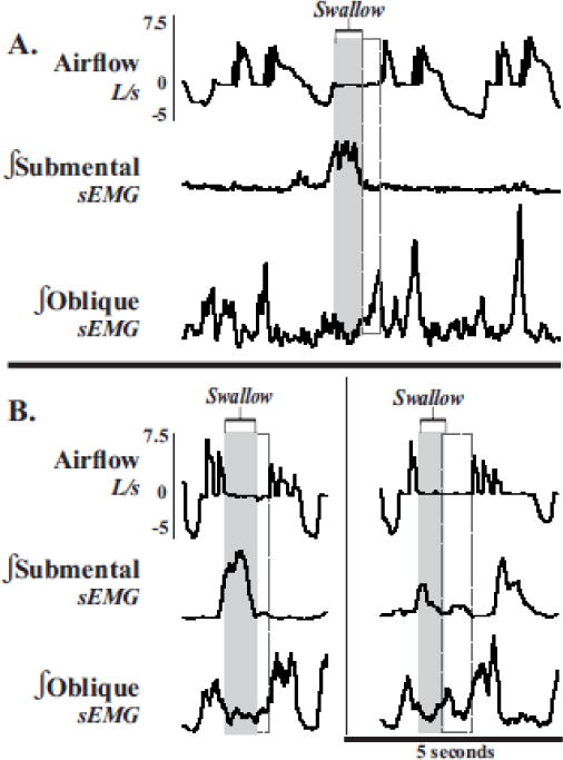
Figure 5. Abdominal EMG depression during compression phases which include swallow.

(A) Integrated submental and abdominal sEMG traces (∫) and airflow measurements show swallow (gray rectangels) and the activation of oblique muscle complex (dashed rectangles), extending the duration of the compression phase. (B, both panels). Of note, this extended compression phase may be due to the need of abdominal muscle activation in preparation for effective shearing forces during the cough expiration.
