Fig. 2.

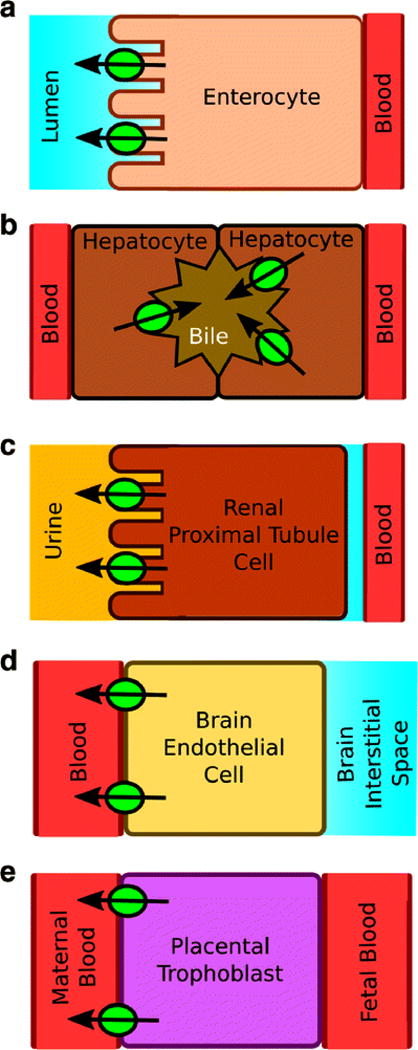
Pgp localization in A) an enterocyte, B) a hepatocyte, C) a kidney proximal tubule cell, D) a brain endothelial cell and E) a placental trophoblast. The green circles are Pgp and the arrows denote the direction of efflux.

Pgp localization in A) an enterocyte, B) a hepatocyte, C) a kidney proximal tubule cell, D) a brain endothelial cell and E) a placental trophoblast. The green circles are Pgp and the arrows denote the direction of efflux.