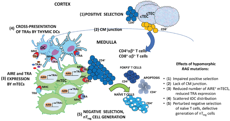
Figure 1. Schematic representation of positive and negative selection in the thymus.
Five steps of T cell maturation and thymocyte selection are shown, which can be affected in patients and animals with hypomorphic RAG mutations. 1) Cortical thymic epithelial cells (cTEC) expressing MHC-peptide ligands mediate positive selection of developing thymocytes. 2) Normal thymic architecture is characterized by demarcation of cortical and medullary areas, with clear evidence of the cortico-medullary junction (shown as dashed dine) where thymic dendritic cells (tDCs) are mainly distributed. Positively selected CD4+ αβ+ and CD8 +αβ+ T cells migrate from the cortex to the medulla, where 3) mature medullary thymic epithelial cells (mTECs) express tissue restricted antigens (TRA) under the control of the transcriptional activator AIRE. CD4+ thymocytes recognizing with high affinity TRAs in the complex with MHC class II molecules on the surface of mTECs or 4) cross-presented by tDCs undergo 5) negative selection or diversion to become FOXP3+ natural regulatory T cells. Hypomorphic RAG mutations may interfere with each of these steps as indicated on the right.

