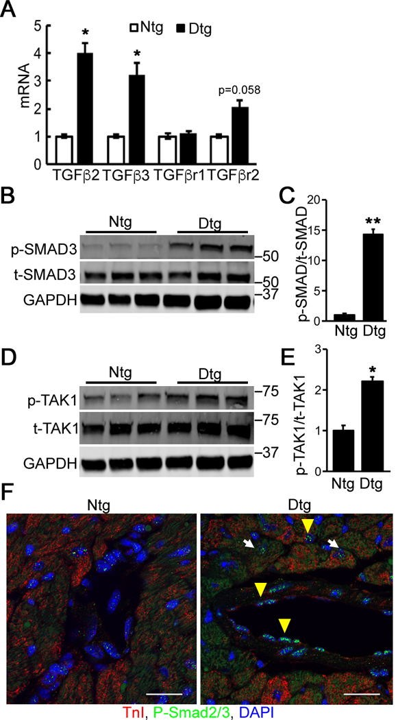
Figure 1. TGFβ signaling is activated in Mybpc340kDa hearts.

A, RNA levels were determined via quantitative RT-PCR for TGFβ ligands and receptors in nontransgenic (Ntg)- and Mybpc340kDa-expressing (Dtg) heart lysates. TGFβ receptor I and II (TβRI and TβRII, respectively), n=3. B, Activation of SMAD3 signaling was detected using Western blot analyses of total cardiac protein. C, The ratio of phosphorylated (p)-SMAD3 to total (t)-SMAD3 was determined from the Western blot, n=4. D, TAK1 levels were determined using Western blot analyses of total cardiac protein. E, The ratio of phosphorylated (p)-TAK1 to total (t)-TAK1 was determined from the Western blot, n=6. F, Immunostaining for p-SMAD2/3 (green) in Ntg and Dtg heart sections derived from the left ventricles. DAPI (blue) was used to detect nuclei and troponin I (red) identified cardiomyocytes. White arrow: p-SMAD2/3 positive cardiomyocyte; yellow arrowhead: p-SMAD3 positive non-cardiomyocyte. Scale bar: 25 μm. Data normalization and between group differences analysis were performed as described in Methods. *P<0.05, **P<0.01. All samples were derived from 4-month-old mice (3 months after withdrawal of doxycycline to activate Mybpc340kDa protein expression).
