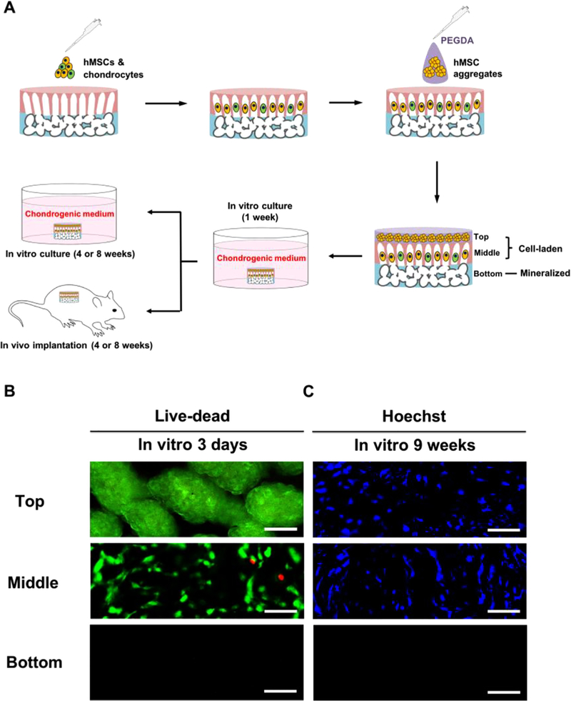
Fig. 2.
The cell-laden trilayer scaffold conduces to stratified cellular alignment after in vitro culture. (A) Schematic for an experimental protocol used to examine tissue-forming ability of the cell-laden trilayer scaffold both in vitro and in vivo. (B) Fluorescent live-dead staining for top, middle, and bottom layers of the cell-laden trilayer scaffold after 3 days of in vitro culture. (C) Fluorescent nucleus (Hoechst) staining for top, middle, and bottom layers of the cell-laden trilayer scaffold following 9 weeks of in vitro culture. Scale bars represent 50 µm.

