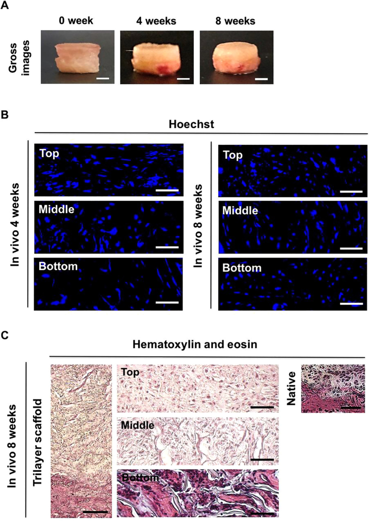
Fig. 3.
The cell-laden trilayer scaffold facilitates the formation of integrated, distinct cartilageand bone-like tissues in vivo. (A) Gross images of the cell-laden trilayer scaffold after 0, 4, and 8 weeks of in vivo implantation. Scale bars indicate 2 mm. (B) Fluorescent nucleus (Hoechst) staining for top, middle, and bottom layers of the cell-laden trilayer scaffold after 4 and 8 weeks of in vivo implantation. Scale bars represent 50 µm. (C) H&E staining of the cell-laden trilayer scaffold following 8 weeks of implantation. Scale bar indicates 200 µm. High magnification images for top, middle, and bottom layers of the trilayer scaffold as well as native osteochondral tissue are also provided. Scale bars represent 50 µm.

