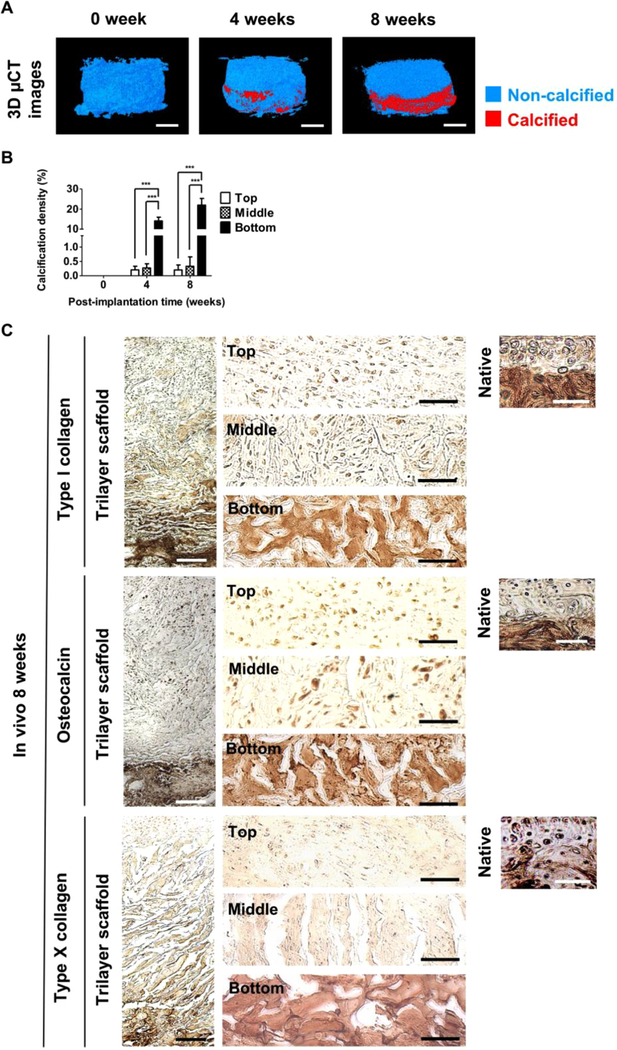
Fig. 4.
The acellular biomineralized bottom layer induces de novo formation of spongy bone in vivo. (A) 3D µCT models of the cell-laden trilayer scaffold after 0, 4, and 8 weeks of in vivo implantation. Calcified and non-calcified tissues are shown in red and blue, respectively. Scale bars indicate 2 mm. (B) Calcification density determined from 3D µCT models of top, middle, and bottom layers of the cell-laden trilayer scaffold as a function of post-implantation time. (C) Immunohistochemical staining for type I collagen, osteocalcin and type X collagen of the cell-laden trilayer scaffold following 8 weeks of implantation. Scale bars indicate 200 µm. High magnification images for top, middle, and bottom layers of the trilayer scaffold as well as native osteochondral tissue are also provided. Scale bars represent 50 µm. Data are shown as mean ± standard errors (n = 6). Comparisons of multiple groups in the same time point were made by one-way ANOVA with Tukey-Kramer post-hoc test. Asterisks indicate statistical significances according to p-values (***p < 0.001). (For interpretation of the references to color in this figure legend, the reader is referred to the web version of this article.)

