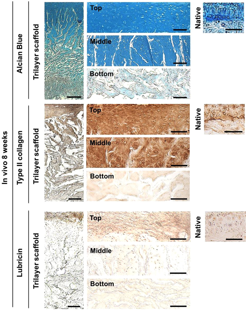
Fig. 5.
The cell-laden top and middle layers support in vivo formation of stratified cartilage with lubricin-rich surface. Alcian Blue staining and immunohistochemical staining for type II collagen and lubricin of the cell-laden trilayer scaffold at 8 weeks post-implantation. Scale bars represent 200 µm. High magnification images for top, middle, and bottom layers of the trilayer scaffold as well as native osteochondral tissue are also shown. Scale bars indicate 50 µm. (For interpretation of the references to color in this figure legend, the reader is referred to the web version of this article.)

