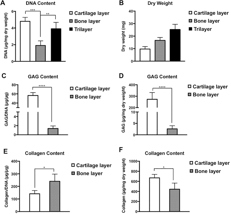
Fig. 6.
Biochemical analyses of the trilayer scaffold. The intact scaffold (Trilayer), its cell-laden top and middle layers (Cartilage layer), and the mineralized bottom layer (Bone layer) were prepared for quantifying the deposition of GAG and collagen after 8 weeks of subcutaneous implantation. (A) DNA content in the scaffolds. (B) Dry weight of the scaffolds. (C, D) GAG content, normalized to the DNA content and dry weight of the corresponding scaffold, respectively. (E, F) Collagen content, normalized to the DNA content and dry weight of the corresponding scaffold, respectively. Error bars show the mean ± SD (n = 8). Statistical significance was performed using one-way ANOVA with post hoc Tukey-Kramer test, *P < 0.1, **P < 0.01, ****P < 0.0001.

