Figure 1.

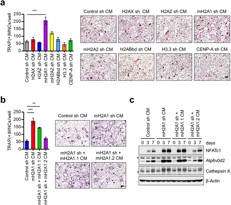
Osteoclastogenic activity of CMs from mH2A1.2-depleted prostate cancer cells. (a) Primary OCP cells derived from adult mouse long bone were cultured with CMs collected from DU145 prostate cancer cells depleted of each of the indicated histone variants in the presence of RANKL. At the end of day 7, OCP-induced cells were fixed, stained for TRAP, and photographed under a light microscope (10×) (right). Representative images of osteoclasts are shown (scale bar, 100 μm). TRAP positive cells containing three or more nuclei and a full actin ring were counted as osteoclasts (left). Cells exposed to CMs from mock-depleted DU145 cells served as control. (b) mH2A1-depleted DU145 cells were infected with lentiviruses expressing shRNA-resistant mH2A1.1 or mH2A1.2, and their CMs were prepared and analyzed for osteoclastogenic activity as in (a). Scale bar, 100 μm. (c) OCP cells were treated with CMs for the indicated time periods, and their lysates were collected and analyzed for the expression of three osteoclast marker genes (NFATc1, Atp6v0d2, and Cathepsin K) by Western blot. β-Actin was used as a loading control. Asterisks indicate non-specific bands. Shown in (a) and (b) are the means ± s.d. (n = 3) of three independent experiments; **P<0.01, ***P<0.001 (ANOVA analysis).
