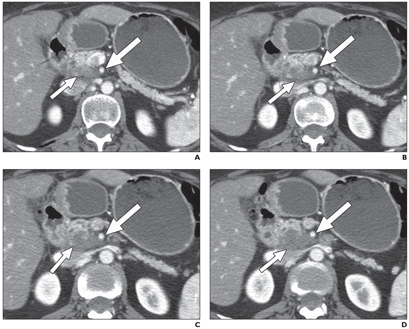
Fig. 1—

55-year-old woman with pancreatic cancer, 3 months after treatment with capecitabine and radiation.
A–D, Contiguous 2.5-mm pancreatic phase axial images. All readers detected 50% or greater contact of low-attenuation uncinate mass (short arrows) with superior mesenteric artery (long arrows), indicating unresectable disease. At surgery, material abutting superior mesenteric artery was inflammatory fibrosis. Patient had R0 resection.
