
Figure 4. Loss of components of anterograde cilia transport induces intracranial hemorrhages in developing zebrafish.
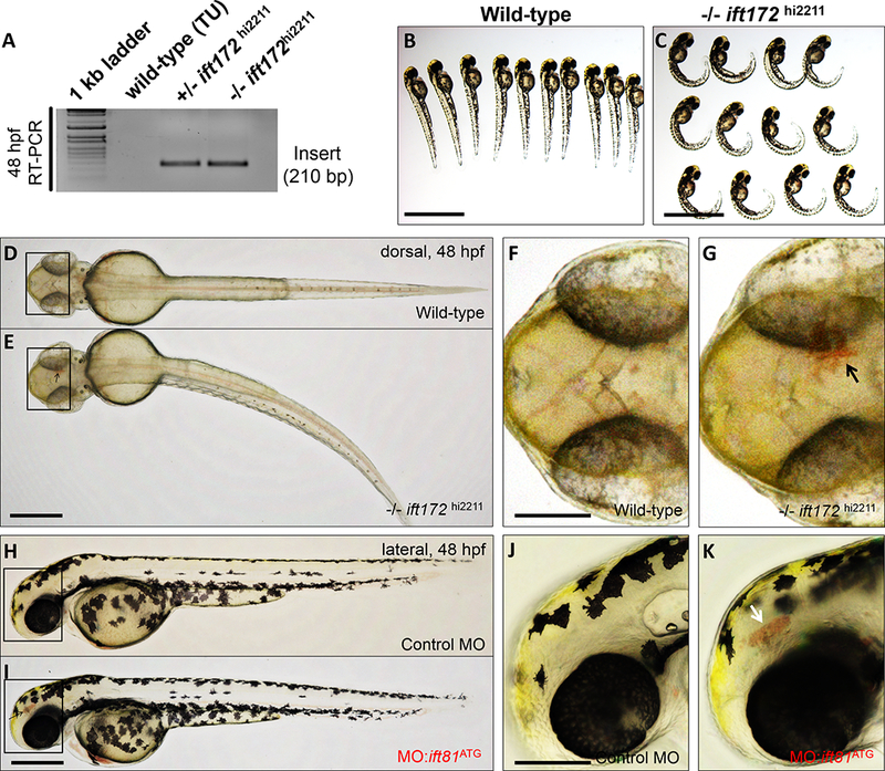
(A) RT-PCR mediated screen for 210 bp viral/insertion-specific amplicon in genomic DNA extracted from wild-type, heterozygous and homozygous ift172hi2211 mutants. (B and C) Bright-field images of representative wild-type and −/−ift172hi2211 mutants at 48–52 hpf are shown. Scale bars: 500 μm. (D and E) Representative dorsal views of whole-mount wild-type and −/−ift172hi2211 zebrafish at 48–52 hpf. Scale bar: 200 μm. (F and G) Higher magnification photomicrographs of the boxed regions in D and E. Black arrow denotes area of hemorrhage in the −/−ift172hi2211 embryo. Anterior is to left. Scale bar: 100 μm. (H and I) Representative lateral views of whole-mount embryos either injected with control morpholino oligonucleotide (control MO) or a morpholino oligonucleotide targeting the ATG site of ift81 (MO:ift81ATG), imaged at 48–52 hpf. Scale bar: 200 μm. (J and K) Higher magnification photomicrographs of the boxed regions in H and I. White arrow points to area of hemorrhage in the MO:ift81ATG-injected embryo. Anterior is to left and posterior to right. Scale bar: 150 μm.