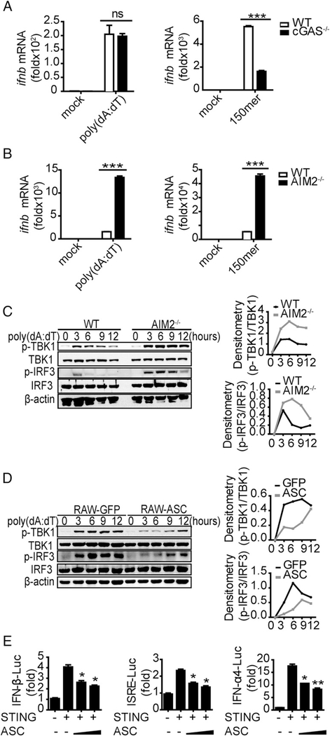
FIGURE 4.
ASC negatively regulates IFN-β production by affecting STING function. (A and B) BMDCs isolated from WT, cGAS−/−, and AIM2−/− mice were stimulated with poly(dA:dT) (1.5 μg/ml) and 150mer dsDNA (1.5 μg/ml), and IFN-β was measured by quantitative real-time PCR (qRT-PCR) 8 h after transfection. Immunoblot of p-TBK1, total TBK1, p-IRF3, total IRF3, and b-actin in WT and AIM2−/− BMDC lysates (C) or RAW-GFP and RAW-ASC cell lysates (D) that were stimulated with poly(dA:dT) (1.5 μg/ml) for the indicated times. (E) Luciferase activity of MEFs that were cotransfected with the IFN-β luciferase reporter (0.4 μg/ml), ISRE luciferase reporter (0.4 μg/ml), or IFN-α4 luciferase reporter (0.4 μg/ml) and Flag-STING (0.4 μg/ml) and the empty vector or increasing amounts of HA-ASC. Data are representative of at least two independent experiments. The results are shown as mean ± SEM. *p < 0.05, **p < 0.01, ***p < 0.001, Student t test. ns, not significant.

