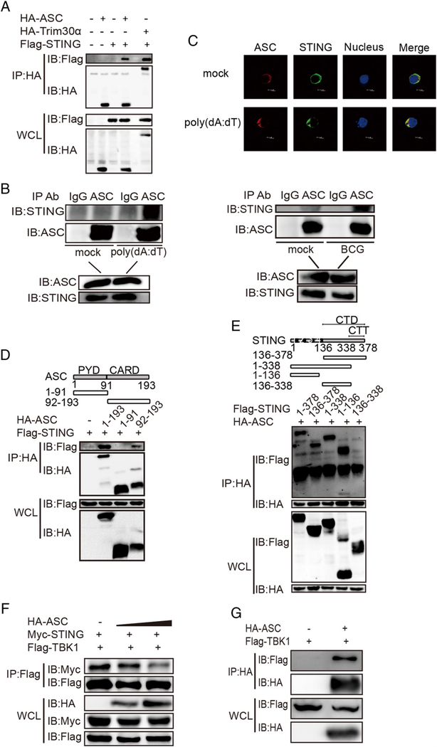
FIGURE 5.
ASC interacts with STING. (A) Immunoblot analysis of lysates from HEK293T cells that were cotransfected with the Flag vector or the Flag-STING vector and the HA-tagged vector, HA-ASC vector, or HA-Trim30a vector, followed by immunoprecipitation (IP) with anti-HA Ab and immunoblotting with anti-Flag Ab. (B) Immunoblot analysis of lysates of BMDCs that were mock treated or stimulated with poly(dA:dT) (1 μg/ml) or infected with BCG (multiplicity of infection = 10) for 6 h, followed by immunoprecipitation with anti-IgG Ab or anti-ASC Ab and immunoblotting with anti-STING Ab. (C) Confocal microscopy of MEFs that were transfected for 4 h with Flag-tagged STING and HA-tagged ASC and then mock treated or stimulated for 4 h with poly(dA:dT) (2 μg/ml). Immunofluorescence was performed using anti-HA Ab (red), anti-Flag Ab (green), and DAPI. (D) Immunoblot analysis of lysates from HEK293T cells that were cotransfected with Flag-STING and HA-tagged ASC or HA-tagged ASC truncations, followed by immunoprecipitation (IP) with anti-HA Ab and immunoblot analysis with anti-Flag Ab. (E) Immunoblot analysis of lysates from HEK293T cells cotransfected with HA-tagged ASC and Flag-STING or Flag-STING truncations, followed by immunoprecipitation (IP) with anti-HA Ab and immunoblot analysis with anti-Flag Ab. (F) Immunoblot analysis of lysates from HEK293T cells cotransfected with Myc-STING and Flag-TBK1 plus the HA vector or HA-ASC in gradient concentrations, followed by immunoprecipitation (IP) with anti-Flag Ab and immunoblot (IB) analysis with anti-Myc Ab. (G) Immunoblot analysis of lysates from HEK293T cells cotransfected with the Flag vector or Flag-TBK1 and HA-ASC, followed by immunoprecipitation (IP) with anti-HA Ab and immunoblot (IB) analysis with anti-Flag Ab. Data are representative of at least two independent experiments.

