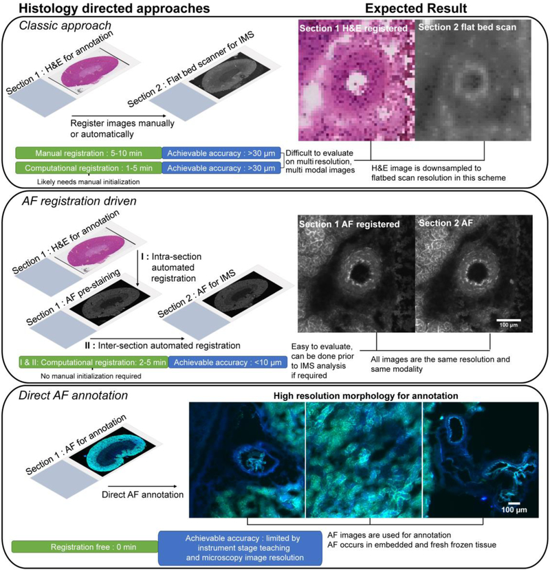
Figure 1. Schematic of histology-driven workflows after tissue sectioning and prior to IMS sample preparation and data acquisition.
Section 1 and 2 refer to serial sections. The AF registration driven result images are after AF-to-AF registration. In the ‘Classic approach’ and ‘AF registration driven’ panels, images were taken from the same region and area for comparative purposes. Colors in the AF scan in ‘Direct AF annotation’ are from a two color channel image using DAPI and FITC filters.

