FIGURE 3.

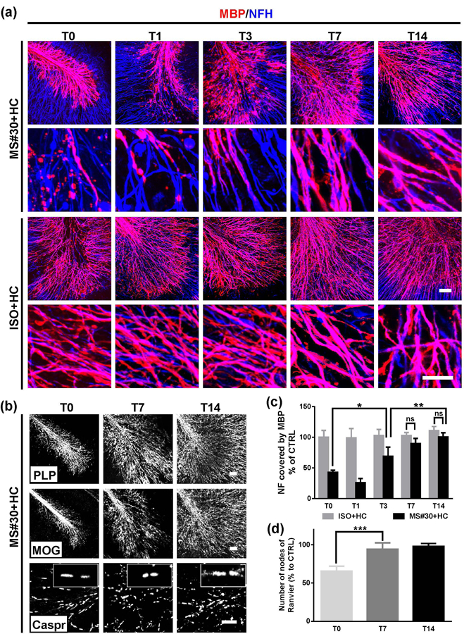
Myelin repair after MS#30+HC demyelination. (a) At the indicated time points after treatment withdrawal, slices were fixed, stained with antibodies against MBP (red) and NF-H (blue), and imaged at low (upper panels) and high (lower panels) magnification using confocal microscopy. (b) Confocal images of slices treated with MS#30+HC and stained with PLP, MOG or Caspr antibodies. (c) The coverage of MBP on NF-H+ axons was calculated at various time points following treatment withdrawal and normalized against control (ISO+HC treated slices) at T0. (d) The number of nodes of Ranvier (paired paranodal Caspar expression) were totaled and then normalized against control (ISO+HC treated slices) at T0. Statistical analyses were performed by unpaired Student’s t test. *: p<0.05, **: p<0.01, ****: p< 0.0001, ns: not significant, n=3–4. Scale bars: 50µm.
