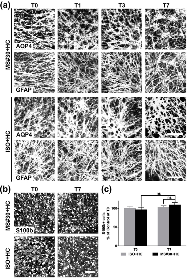
FIGURE 4.

Number and morphology of astrocytes following MS#30+HC treatment. (a) Confocal images of cerebellar slices treated with either MS#30 or ISO plus HC and stained for (a) AQP4 and GFAP or (b) S100b. (c) quantification of S100b staining in slices. No significant difference of S100b+ cells at T0 and T7 in MS#30+HC treatment or between MS#30+HC and ISO+HC. Statistical analyses were performed by multiple unpaired Student’s t test. ns: not significant, n=3–4. Scale bars: 50µm.
