Fig. 4.
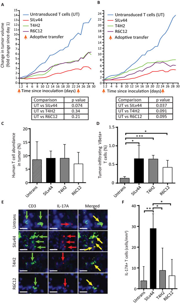
TCR-transduced T cells differentially contain melanoma tumors. (A) SCID/beige mice were subcutaneously injected with 106 888-A2+ human melanoma tumor cells at three mice per group and allowed to firmly establish tumors over three weeks, before 2×10⁶ human T cells were adoptively transferred twice with a 23 day interval. Tumor growth was significantly contained only by SILv44 transduced T cells. (B) The experiment was repeated at five mice per group and again, only the SILv44 TCR transduced T cells significantly contained tumor growth. (C) Intratumoral T cells were equally abundant at euthanasia regardless of the expressed TCR, showing that it was T cell function rather than T cell number holding tumors in check. (D) Transgenic TCR expression (as % of tumor infiltrating T cells) was limited to transduced T cell groups as determined using antibodies to the TCRβ subunit. (E) An increased frequency of IL-17A+ T cells was found in tumors infiltrated by SILv44 expressing T cells. (F) Immunohistology was performed and quantified on multiple sections of two tissue samples per group. Scale bars represent 17µm. *P<0.05, ** P<0.1, *** P<0.001.

