
Figure 1.
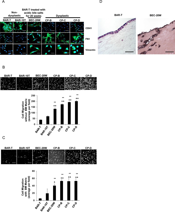
Non-dysplastic Barrett’s cells treated with 20 weeks of acidic bile salt exposure (BEC- 20W) demonstrate features of EMT. (A) Immunofluorescent staining for CDH1, FN1, and vimentin. Cell migration without (B) and with (C) a basement membrane (BM) matrix. (D) Cell migration in 3D organotypic culture. Scale bars = 20 µm. For cell migration assays in B &C, bar graphs depict the mean ± SEM of three independent experiments. **p<0.01 vs. BAR-T; ++p<0.01 vs. BAR-10T.