
Figure 2.
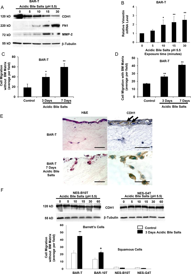
Acidic bile salts induce features of EMT in non-neoplastic Barrett’s (BAR-T) cells, but not in non-neoplastic esophageal squamous (NES) cells. (A) Western blot for CDH1, FN1, and MMP-2; β-tubulin served as a loading control. (B) Representative qPCR for vimentin mRNA level after acidic bile salt treatment relative to untreated (0 minutes) cells; qPCR assays were performed in triplicate in at least two independent experiments. Cell migration after 3 and 7 days of acidic bile salt exposure without (C) and with (D) a basement membrane (BM) matrix. (E) Cell migration and CDH1 immunostaining of BAR-T cells grown in 3D organotypic culture after 7 days of acidic bile salts. Arrows depict membranous CDH1 staining. Scale bars = 20 µm. (F) Western blot for CDH1 and cell migration in NES cells. Bar graphs depict the mean ± SEM, *p<0.05 and **p<0.01 compared with 0 minutes or control; +p<0.05 compared with BAR-10T control. For cell migration assays in C, D, & F, bar graphs depict the mean ± SEM of three independent experiments.