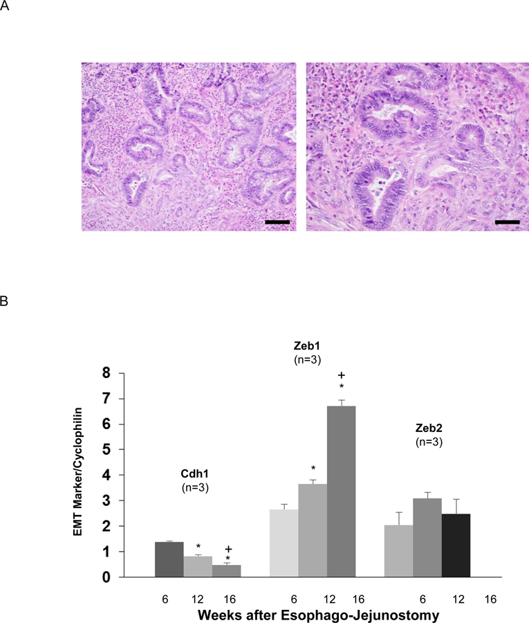
Figure 4.

Columnar-lined esophagus (CLE) that develops in rats with surgically-induced reflux esophagitis demonstrates markers of EMT. (A) H&E staining of neoglandular epithelium at medium power (Scale bar = 100 µM) in the left panel and high power (Scale bar = 50 µM) in the right panel (B) Representative qPCR for Cdh1, Zeb1, and Zeb2 mRNA in CLE at week 6, 12, and 16 post-operatively. For each mRNA, relative levels were normalized to cyclophilin. qPCR assays were performed in triplicate in 3 individual animals. Bar graphs depict the mean ± SEM. *p<0.05 compared with week 6; +p<0.05 compared with week 12.
