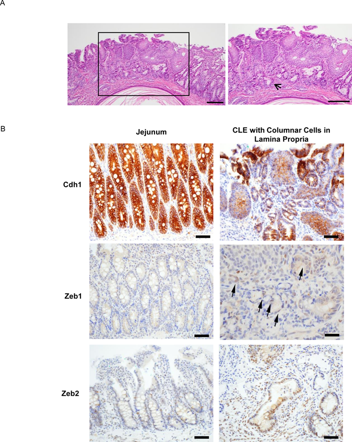
Figure 5.

In a rat model of reflux esophagitis, CLE with columnar cells in the lamina propria underneath esophageal squamous epithelium demonstrates markers of EMT. (A) H&E staining of CLE (bracketed region) in the distal esophagus. Right panel showed the enlarged bracketed region, indicating extensive subsquamous glands (arrow; scale bar = 200 µM). (B) Immunostaining for Cdh1, Zeb1, and Zeb2 in native jejunal epithelium (left side of each pair) and CLE with columnar cells in the lamina propria (right side of each pair). Arrows indicate columnar cells in the lamina propria with Zeb1 expression. All scale bars = 50 µM except for Zeb1 whose scale bar = 25 µM.
