Figure 7.

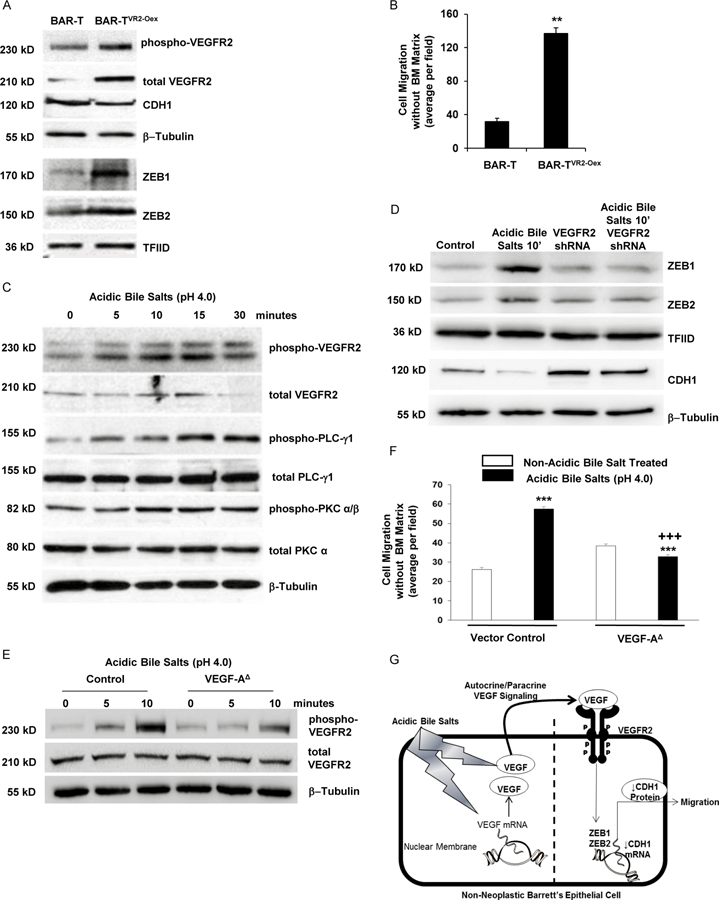
Acidic bile salts-induce VEGF/VEGFR2 signaling that mediates EMT features in non- neoplastic Barrett’s cells. (A) Western blot for phopho-VEGFR2, CDH1, ZEB1, and ZEB2 in BAR-T cells with and without VEGFR2 overexpression (VR2-Oex) and (B) cell migration in BAR-T cells with and without VEGFR2 overexpression. Bar graphs depict the mean ± SEM of three independent experiments. **p<0.01 compared with control. (C) Western blot for phopho- VEGFR2 and its downstream pathway proteins phospho-PLC-γ1 and –PKC α/β after exposure to acidic bile salts. (D) Western blot of ZEB1, ZEB2 and CDH1 with and without VEGFR2 knockdown by shRNA following acidic bile salt exposure; empty vector-containing cells served as control. β-tublin and TFIID served as loading controls for whole cell and nuclear lysates, respectively. (E) Western blots for phospho-VEGFR2 with and without VEGF-A deletion (VEGF- A∆) following acidic bile salt exposure; empty vector-containing cells served as control. (F) Cell migration in BAR-T cells with VEGF-A∆ following acidic bile salt exposure. Bar graphs depict the mean ± SEM of three independent experiments. ***p<0.001 compared with corresponding control; +++p<0.001 compared with acidic bile salt treated vector control. (G) Schematic model demonstrating the EMT-inducing effects of acidic bile salts in non-neoplastic Barrett’s cells. Exposure to acidic bile salts causes a rapid increase in VEGF secretion along with a delayed increase in VEGF mRNA expression. The secreted VEGF binds in an autocrine/paracrine fashion to VEGFR2, resulting in increased expression of ZEB1 and ZEB2, which decrease the expression of CDH1 and increase cell migration.
