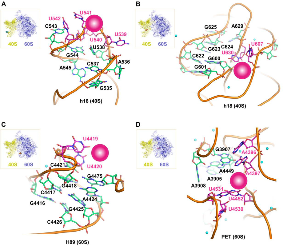
Figure 3. Modeled structural representations of putative binding sites for the tetracyclines Col-3 and doxycycline.
Modeled human ribosomal sites showing potential rRNA binding pockets for Col-3 and doxycycline on the small, 40S subunit at (A) h16 and (B) h18. Modeled sites showing potential rRNA binding pockets for Col-3 and doxycycline on the large, 60S subunit at (C) H89 (D) peptidyl exit tunnel (PET). Crosslinked bases are highlighted in magenta and magenta spheres represent potential binding pockets for Col-3 and doxycycline at each site. Small green spheres represent divalent magnesium ions.

