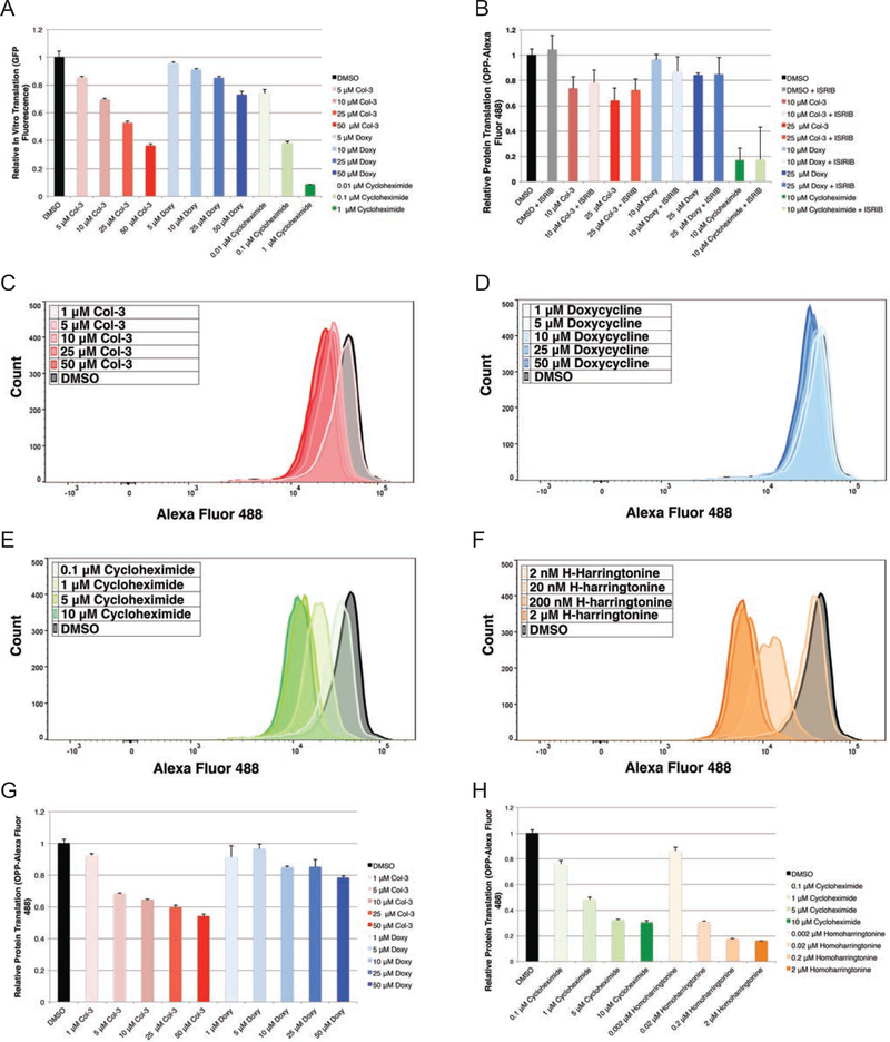
Figure 5– IVT and OPP-flow cytometry analysis shows tetracyclines inhibit human translation independent of activation of the ISR.
(A) Bar graph showing relative in vitro translation (IVT) of a GFP reporter on tetracycline- and cycloheximide-treated human ribosomes after 6 h (B) Bar graph showing relative incorporation of O-propargyl puromycin into nascent peptides from tetracycline-treated A375 cells as measured by mean fluorescence (Alexa Fluor 488) in A375 cells co-dosed with tetracyclines ± 200 nM ISRIB at 3 h. Histogram plots for relative incorporation of OPP-Alexa Fluor 488 into nascent peptides in K562 cells dosed for 3 h at multiple concentrations with (C) Col-3 (D) doxycycline (E) cycloheximide and (F) homoharringtonine. (G) Bar graphs showing relative incorporation of OPP-Alexa Fluor 488 into nascent peptides in K562 cells dosed for 3 h with Col-3, doxycycline, cycloheximide, and homoharringotnine. Data are represented as mean ± SD. IVT and flow cytometry data represent experiments with a minimum of three independent replicates. See also Figure S4.

