
Fig. 5. Co-localization of A3A and A3B with HBV core protein (HBc).
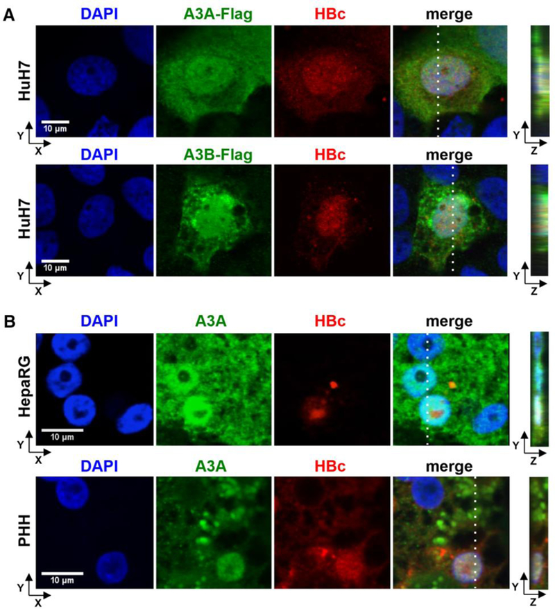
(A) HuH7 cells were co-transfected with an HBV1.1-fold genome and A3A-Flag or A3B-Flag expressing plasmids and stained using DAPI, anti-HBc and anti-FLAG antibodies. (B) HBV-infected dHepaRG and PHH were treated with IFN-α at day 7 post infection for 3 days. A3A and HBc were analyzed by immunofluorescence staining. Right panels indicate z stacks taken at the dotted lines.