Fig. 1.
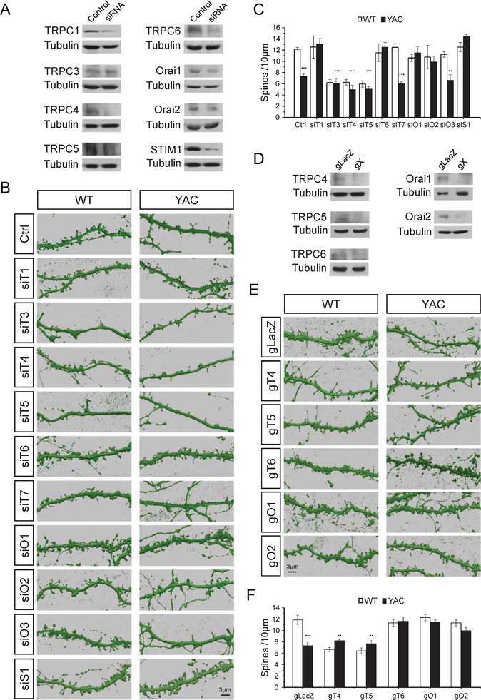
MSN spine density measurements in WT and YAC128 corticostriatal cultures. (A, D) Western blots of lysates from corticostriatal co-cultures after lenti-viral infection of neurons with siRNA (A) or Cas9/gRNA (D) targeting nSOC pathway components. The X in gX represents the indicated subunit. (B, E) 3D reconstructions from confocal images of DARPP32-immunostained MSN dendrites. Spines are shown for DIV 20 corticostriatal co-cultures prepared from WT and YAC128 pups. Co-cultures were infected with lentiviruses to express scrambled siRNA (Ctrl) or siRNA targeting TRPC1–7 (siT1-siT7), Orai1–3 (siO1-siO3), or STIM1 (siS1) (B). Co-cultures infected with lentiviruses to express Cas9 and control sgRNA (gLacZ), sgRNA targeting TRPC4–6 (gT4, gT5, gT6), or Orai1–2 (gO1, gO2) (E). (C, F) Quantification of spine density per 10μm of dendritic length in WT and YAC128 cultures for RNAi knockdown experiments (C) and CRISPR/Cas9 knockout experiments (F). The results are shown as mean±S.E. (n=5–10). *p<0.05, **p<0.01, ***p<0.001 when compared to spine density in WT control cultures.

