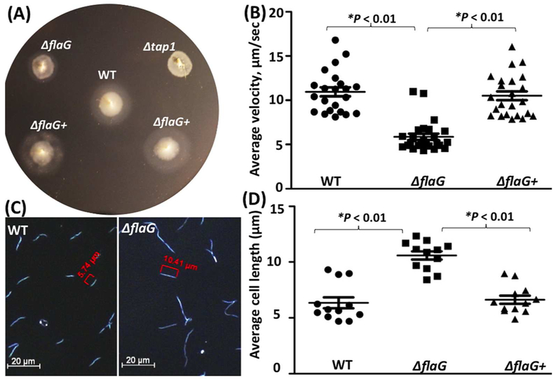
Figure 4. Deletion of flaG alters the cell length and motility.
(A) Swimming plate assay of the wild-type, ΔflaG, and ΔflaG+ strains. The assay was carried out on 0.35% agarose containing the TYGVS medium diluted 1:10 with PBS. The plates were incubated anaerobically at 37°C for 5 days. Δtap1, a non-motile mutant, was used as a control to determine initial inoculum sizes. (B) Cell-tracking analysis of the wild-type, ΔflaG, and ΔflaG+ strains. The cells were tracked in the presence of 1% methylcellulose as previously described. The results were the average of at least 20 cells and are expressed as the mean of μm/s ± standard errors of mean (SEM). (C) Dark-field microscopic images of the wild-type and ΔflaG strains. The cells were visualized under dark-field illumination at 20× magnification using a Zeiss Axiostar plus microscope. (D) Average cell lengths of the wild-type, ΔflaG, and ΔflaG+ strains. The results were the average of at least 12 cells and expressed as the mean of μm ± SEM. The data were analyzed by one-way ANOVA followed by Tukey’s multiple comparison at P < 0.01.

