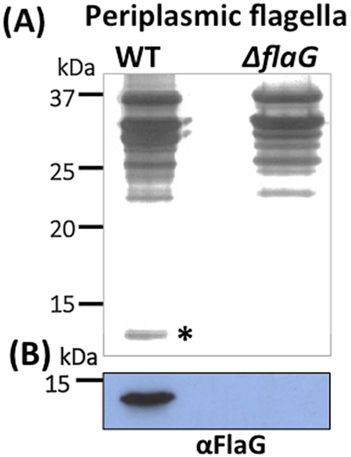
Figure 7. FlaG was detected in the isolated PFs.
(A) SDS-PAGE analysis of the PFs isolated from the wild-type and ΔflaG strains, followed by silver staining. Asterisk points to FlaG. (B) Immunoblotting analysis of the PFs isolated from the wild-type and ΔflaG strains with a specific antibody against FlaG (αFlaG).

