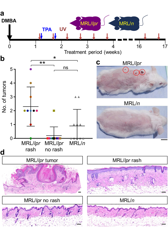
Figure 2. Cutaneous lupus inflammation promotes skin cancer development.

(a) Schematic diagram of the protocol designed to test the role of cutaneous lupus inflammation in skin cancer promotion. All animals received DMBA/TPA to initiate skin carcinogenesis followed by chronic UV exposure in order to induce and maintain skin rash in MRL/lpr mice. (b) Number of tumors developed in MRL/lpr mice on the back skin with rash (upper back), back skin with no rash (lower back) and MRL/n control mice are shown. Colored points highlight the areas of rash and no rash in the same MRL/lpr mouse (*p < 0.05; **p < 0.01 by Student’s t test). (c) Representative images of MRL/lpr and MRL/n mice following skin carcinogenesis protocol are shown. Red circles highlight skin tumors on MRL/lpr skin with rash; scale bar: 1 cm. (d) Representative histological images of skin tumor, skin rash and skin with no rash of MRL/lpr and back skin of MRL/n mice after the completion of skin carcinogenesis protocol are shown; scale bars: 100 µm. Age-matched female mice were used in this study; n = 10 per group.
