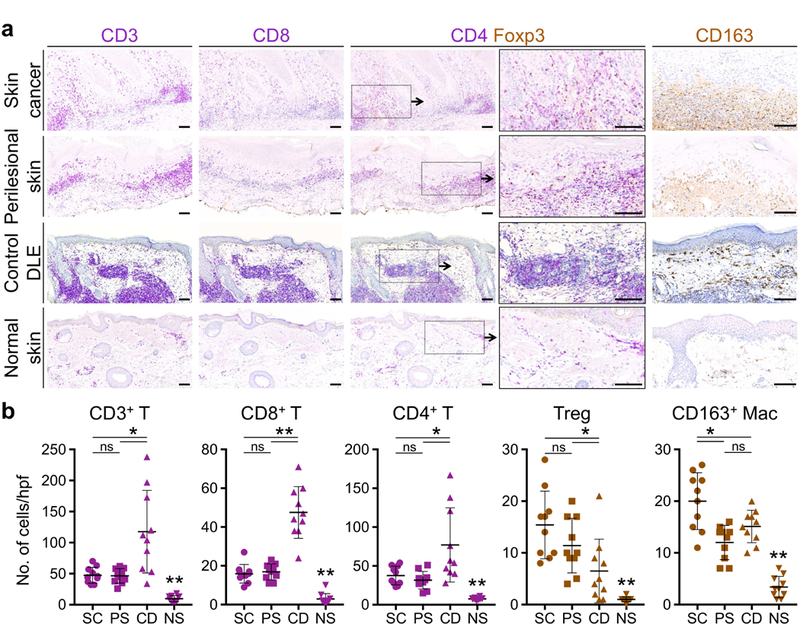
Figure 5. Patient’s DLE plaque margins are infiltrated by tumor-promoting immune cells surrounding the sites of skin cancer development.

(a) T cell and macrophage accumulation in the skin cancer and perilesional skin of cancer-prone DLE lesion is compared to DLE lesions from three cancer-free African American patients (control DLE) and an age-matched normal facial skin as shown by representative images of CD3, CD8, CD4/Foxp3 and CD163-stained tissue sections. (b) Average number of CD3+ T, CD8+ T, CD4+ T, Foxp3+ Tregs and CD163+ M2 macrophage are determined by counting the cells in 10 random hpf of the skin cancer (SC), perilesional skin of DLE (PS), normal skin (NS) excision sections, and 10 random hpf across punch biopsy specimen from three independent control DLE lesions (CD; Supplementary Figure 10). Stained cells were counted blindly; ns: not significant, *p < 0.05, **p < 0.0001 by Student’s t test; scale bars: 100 µm.
