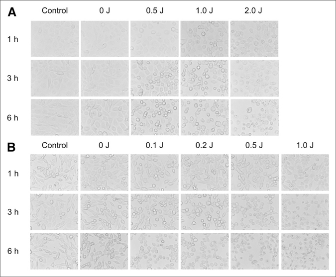
FIGURE 1.
In vitro microscopy studies on A431 cells (A) and 3T3/HER2 cells (B) after photoimmunotherapy. In both cell lines, excitation light induced cellular swelling, bleb formation, and rupture of vesicles representing necrotic cell death. Change of cell morphology correlated with dose of light and also increased over time after photoimmunotherapy.

