FIGURE 4.
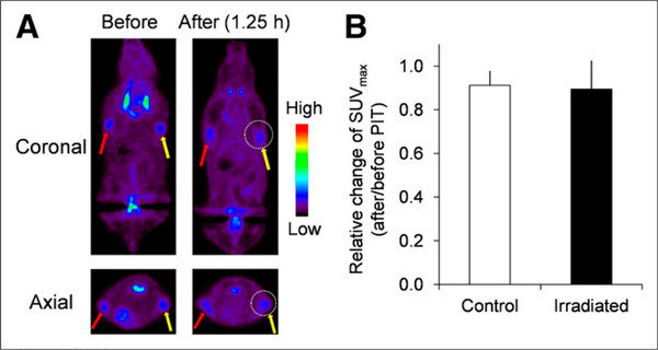
In vivo 18F-FDG PET imaging before and after photoimmunotherapy in target-negative animal model. (A) 18F-FDG PET images were acquired 1.25 h after photoimmunotherapy with panitumumab-IR700 in Balb3T3/DsRed tumor (HER1-negative)–bearing mice. (B) Quantitative data analysis on relative change of SUVmax (after and before photoimmunotherapy). Data are represented as mean ± SEM (n= 4–6 mice). Uptake of 18F-FDG was not changed before and after photoimmunotherapy. PIT = photoimmunotherapy.

