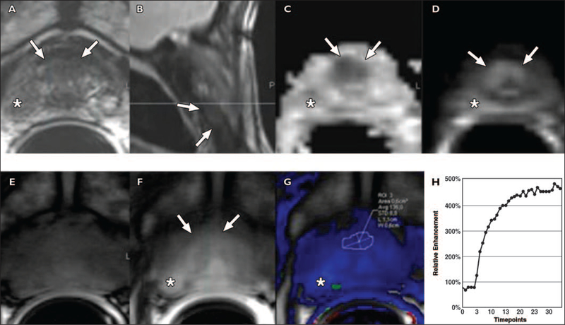
Fig. 9 —
Anterior hypovascular central gland carcinoma in 68-year-old man with prostate-specific antigen level of 11 ng/mL. One set of 12 transrectal ultrasound (TRUS)- guided prostate biopsy results was negative. Images show focal right nonspecific hypointense signal (asterisk, A) with no significant restriction of diffusion (asterisk, C and D), and no washout on color-coded quantitative dynamic contrast-enhanced (DCE) map (asterisk, F and G). Four of four positive anterior biopsies directed within anterior hypointense signal yielded prostate adenocarcinoma, Gleason score of 6, cancer length of 24 mm, negative biopsies in right peripheral zone and in 12 TRUS-guided prostate biopsies.
A and B, Axial (A) and sagittal (B) T2-weighted MR images show anterior apical homogeneous ill-defined lenticular hypointense signal (arrows), suggesting central gland tumor.
C and D, Low apparent diffusion coefficient value (C) and bright signal intensity at b1000 diffusion-weighted (D) images suggest malignancy (arrows).
E and F, Unenhanced (E) and qualitative DCE (F) MR images show no significant enhancement in anterior focal area (arrows, F).
G and H, Low-color-coded kep (reverse reflux rate constant) value (G) and type 1 semiquantitative DCE curve (8.5 s/timepoint) (H) suggest benign tissue.

